The extracellular juncture domains in the intimin passenger adopt a constitutively extended conformation inducing restraints to its sphere of action.

Weikum J, Kulakova A, Tesei G, Yoshimoto S, Jægerum LV, Schütz M, Hori K, Skepö M, Harris P, Leo JC, Morth JP, Sci Rep 10(1):21249 (2020) Europe PMC

SASDJ84 – Intimin D00-D0 domain

Intimin, D00-D0 domain (6xHis tagged)
MWexperimental 21 kDa
MWexpected 23 kDa
VPorod 32 nm3
log I(s) 4.21×10³ 4.21×10² 4.21×101 4.21×100
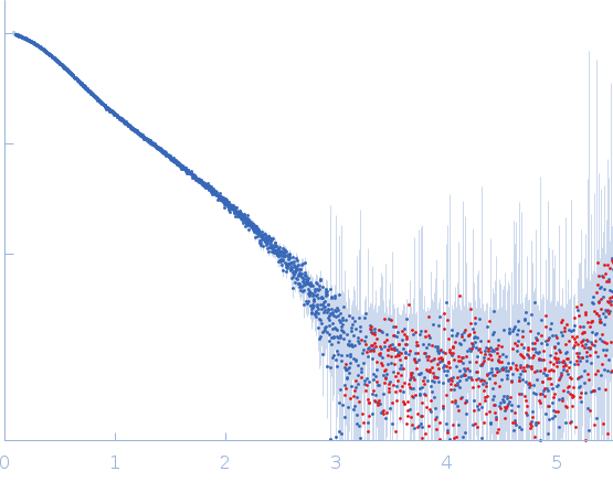
Intimin, D00-D0 domain (6xHis tagged) small angle scattering data  s, nm-1
ln I(s)
Intimin, D00-D0 domain (6xHis tagged) Guinier plot ln 4.21×10³ Rg: 3.1 nm 0 (3.1 nm)-2 s2
(sRg)2I(s)/I(0)
Intimin, D00-D0 domain (6xHis tagged) Kratky plot 1.104 0 3 sRg
p(r)
Intimin, D00-D0 domain (6xHis tagged) pair distance distribution function Rg: 2.7 nm 0 Dmax: 8.3 nm

Data validation


Fits and models


log I(s)
 s, nm-1
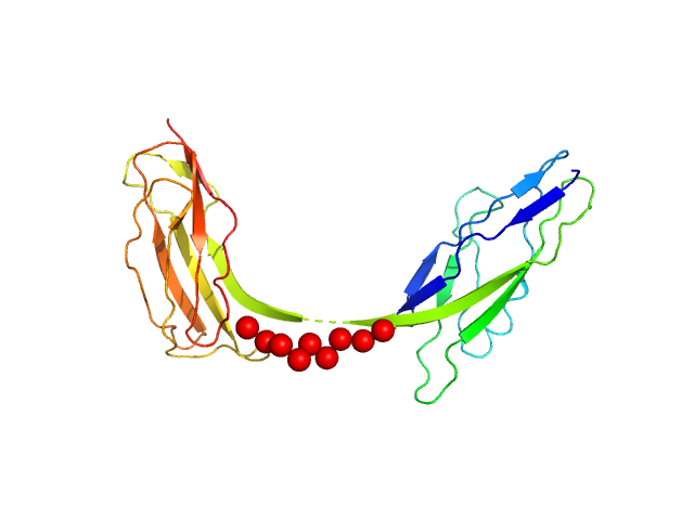
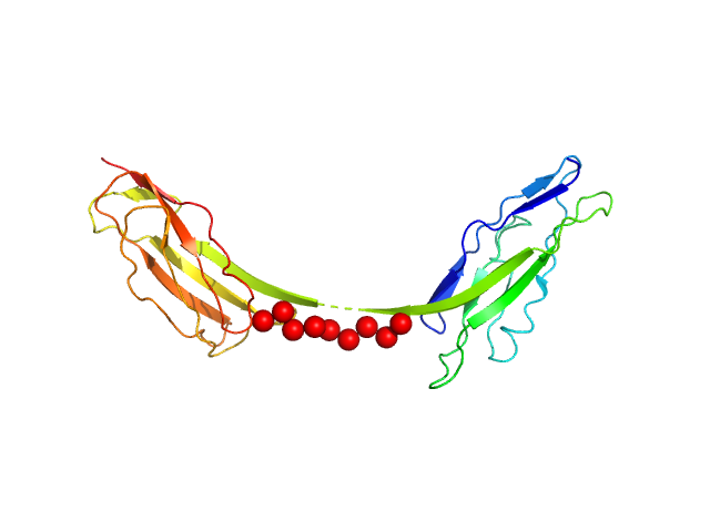
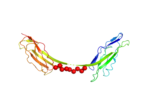
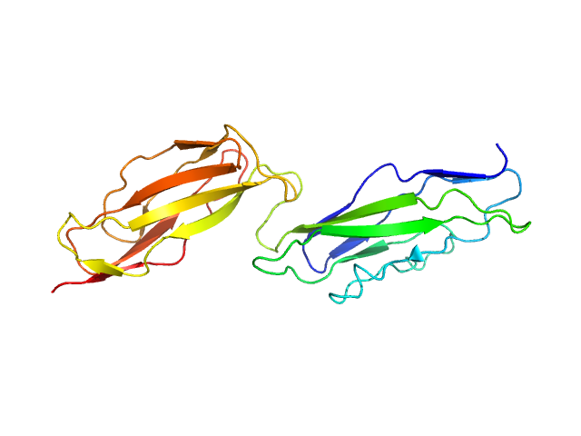
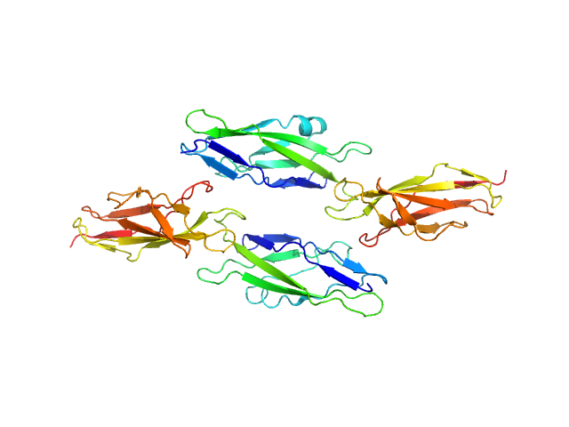
Intimin, D00-D0 domain (6xHis tagged) EOM/RANCH model

log I(s)
 s, nm-1
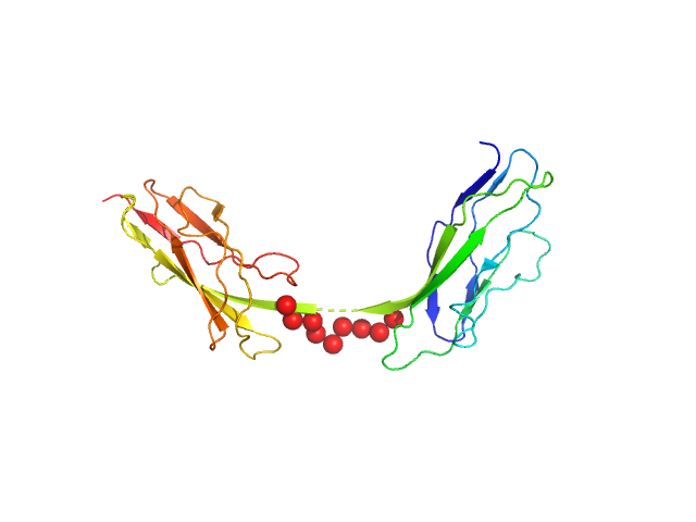
Intimin, D00-D0 domain (6xHis tagged) EOM/RANCH model
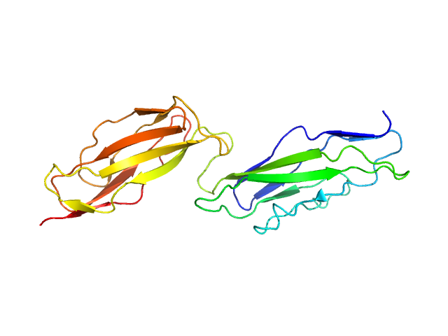
Intimin, D00-D0 domain (6xHis tagged) PDB (PROTEIN DATA BANK) model

log I(s)
 s, nm-1
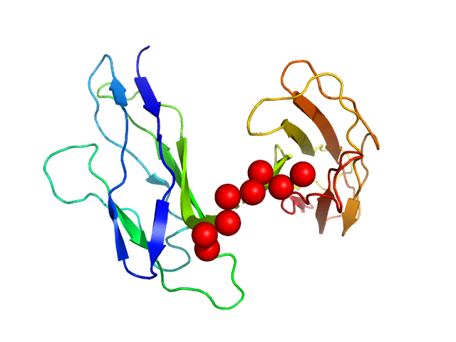
Intimin, D00-D0 domain (6xHis tagged) EOM/RANCH model
Intimin, D00-D0 domain (6xHis tagged) PDB (PROTEIN DATA BANK) model

Synchrotron SAXS data from solutions of the intimin D00-D0 domain in 10 mM HEPES, pH 7.5 were collected on the EMBL P12 beam line at PETRA III (DESY, Hamburg, Germany) using a Pilatus 6M detector at a sample-detector distance of 4 m and at a wavelength of λ = 0.123981 nm (I(s) vs s, where s = 4πsinθ/λ, and 2θ is the scattering angle). Solute concentrations ranging between 1 and 11 mg/ml were measured . 20 successive 0.195 second frames were collected. The data were normalized to the intensity of the transmitted beam and radially averaged; the scattering of the solvent-blank was subtracted. The low angle data collected at lower concentration were merged with the highest concentration high angle data to yield the final composite scattering curve.

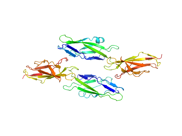
The buffer measured before and the buffer measured after the sample measurement did not match. Therefore, averaged data for buffer and for sample were scaled at high s (starting from point nr 2000), resulting in the high noise at the high s. All data analysis was focused on the low-s region (up to s=0.2 nm^-1). The models represent the ensemble and oligomeric states of the protein in solution at various sample concentrations (top = 6 mg/ml; middle = 1 mg/ml; bottom = 11 mg/ml). The concentration series data are made available in the full entry zip archive.

Intimin, D00-D0 domain (6xHis tagged) (D00-D0)
Mol. type   Protein
Organism   Escherichia coli O127:H6 (strain E2348/69 / EPEC)
Olig. state   Monomer
Mon. MW   22.9 kDa
 
UniProt   P19809 (450-650)
Sequence   FASTA
 
PDB ID   6TQD